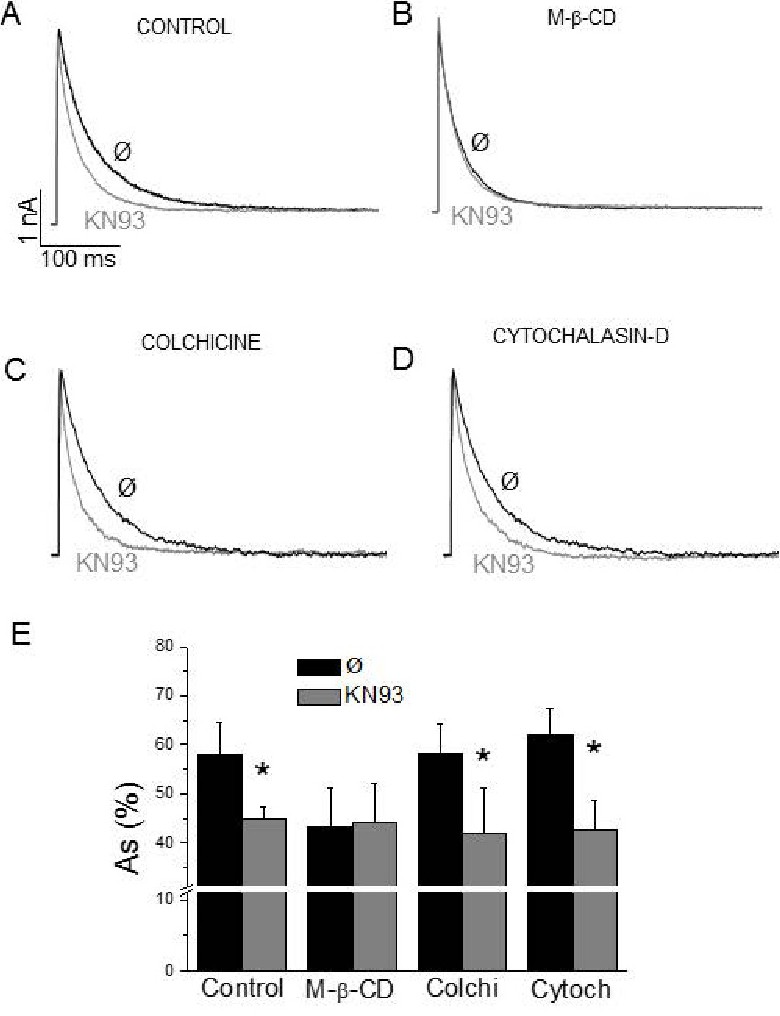
Fig. 1. CaMKII slows down the Ito inactivation kinetics only in intact non-caveolar membrane rafts. Superimposed Ito recordings from two representative myocytes: one recorded in control conditions (Ø) and the other one with KN93 in the internal solution. A) Myocytes in control conditions, B) Myocytes lacking membrane rafts after M-β-CD incubation, C) Myocytes lacking caveolae due to incubation with colchicine or D) Cytochalasine-D. E) Relative contribution of the slow component of Ito inactivation (As) in the same experimental groups. n=6-10. *p<0.05 (compared to control).